Mammals necropsy
External examination
It begins with the external inspection of the carcass, which should collect data concerning:
- Body condition (emaciated, thin, good condition, overweight, obese). It can be evaluated on a scale of 1 to 5.
- The degree of hydration by assessing the skin fold and the sinking of the eyeballs.
- The presence of skin lesions (alopecia, erythematous areas, wounds, abscesses, etc.) and examination of the appendages (hooves, horns, nails, pads).
- Examination of the color of the mucosa of the natural openings (oral, ocular, anal, penile and vaginal). It can be pale pink, indicating anemia or hypovolemia; blue (cyanotic) due to sepsis, dark-red (congestive) due to circulatory alterations, or yellow (icteric) due to hepatic diseases.
- In addition, it will be taken the opportunity to evaluate at the same time in the oral cavity the bone conformation (brachygnathia, prognathism, malocclusion, etc.), as well as the gums, teeth, tongue and palate to determine the presence of lesions (papules, vesicles, bullae, erosions, or ulcers). The study of the teeth can help to estimate the age of the animal according to the morphology, and the lack or presence of certain teeth. In addition, it allows to evaluate the type of feeding of the animal.
- Inspection of the external palpable lymph nodes (size, consistency, etc.).
- Inspection of the umbilical region and joints (especially in young animals).
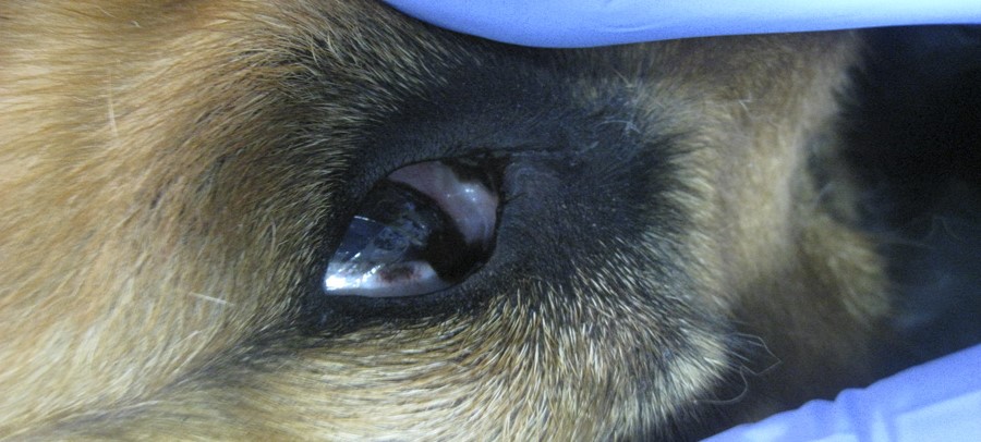
Mammal necropsy. External examination
Internal examination:
PSubsequently, the carcass is inspected internally. Once at this point, it is very important to follow an established order so as not to forget to inspect any part. However, in some circumstances, due to some specific characteristic of the animal or the pathological process, it may be necessary to make some modification in the technique.
It should bear in mind that the study of the organs should be carried out immediately after their extraction, except for the digestive tract, which should be done in the last place, since it can contaminate the rest of the samples and tissues. To explore the tubular organs (digestive tract) a longitudinal opening should be performed, and in the case of the solid/parenchymatous organs, filleting is recommended.
The animal should be placed in right lateral recumbence, that is to say, the right side of the animal has to be supported on the table/surface where the necropsy will be performed.
The left forelimb is dismembered, at axillary level, cutting the muscles, as well as the left hind limb, at the level of the coxofemoral joint, leaving both limbs attached to the carcass by their dorsal portion. Taking advantage of this step, the preescapular and subscapular lymph nodes are localized and examined.
A longitudinal skin incision is made from the submandibular space to the perineum following the midline of the abdomen and then separating the skin from the subcutaneous connective tissue until reaching the paravertebral area. At this point, the coloration, degree of hydration, possible trauma, hematomas, etc should be evaluated. In the case of male animals and in mares and adult female ruminants, the penis or udder is separated by means of cuts around these organs. The mammary gland is examined by palpation and transverse cuts, and the testicles by a longitudinal incision that will affect both the testicular parenchyma and the epididymis.
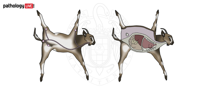
Necropsy in the goat. Positioning in left lateral decubitus. Skin incision lines and opening of the abdominal and thoracic cavities.
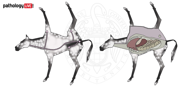
Necropsy in the horse. Positioning in right lateral decubitus. Skin incision lines and opening of the abdominal and thoracic cavities.
To open the abdominal cavity, three cuts should be made in the musculature of the left abdominal wall: one from the xiphoid appendix to the symphysis pubis, another caudal to the costal arch from ventral to dorsal and the last one at the level of the groin. Once the musculature is removed, it should be assessed both the presence of abnormal fluids (coloration, quantity, density) and the topography of the abdominal organs.
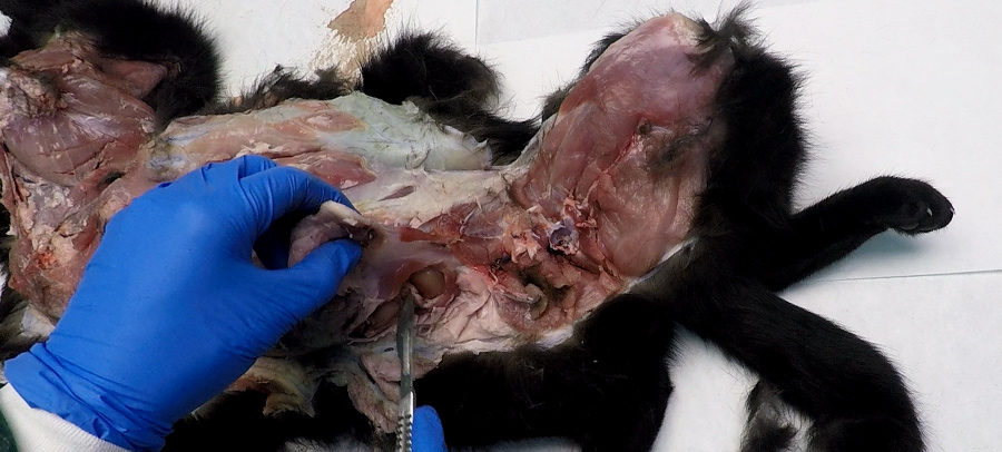
Opening of the abdominal cavity
At this point, it is very important to make a cut with the scalpel blade in the diaphragmatic dome, to check if the thoracic cavity maintains the characteristic negative pressure. If so, air will enter the thorax, leading to caudal displacement of the diaphragm, emission of a characteristic sound and collapse of the healthy lungs.
The opening of the thorax will be performed as follows: with a scalpel or a knife, a cut is made from the last rib to the first rib, as close as possible to the spine (dorsal part) and the sternum (ventral), cutting superficial muscles. Then, proceed to cut the ribs with the costotome following the drawn line. At this point, the ribs are inspected, assessing the consistency and the costochondral junction (degree of ossification, bone growth line and examination of the bone marrow). Once the ribs are removed, the presence of abnormal contents and the position of the thoracic organs are evaluated.
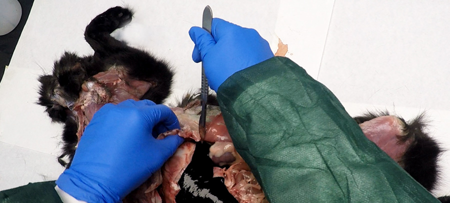
Opening of the thoracic cavity
In addition, a small incision is made in the pericardial sac at the level of the apex to assess the presence of contents inside (fluid, quantity, coloration, presence of fibrin, etc.).
The pelvic cavity is opened by making two cuts, with a saw or costotome, on each side of the symphysis pubis, taking advantage of the obturator holes through the pubis and the ischial arcade. This step will allow the removal of the pelvic floor in order to later be able to completely extract the abdominal organs.
In order to carry out the examination of the organs, they should be extracted from the cavities; first the abdominal ones (the digestive apparatus together with the spleen, liver, pancreas and the urinary and genital apparatus) and then the thoracic ones (tongue, esophagus, larynx, trachea, lungs and heart).
Abdominal organs
At the level of the diaphragm (caudal or cranial to the diaphragm, the esophagus has to be ligated to avoid the exit of stomach or esophageal contents, with the consequent contamination of the organs). The gastrophrenic and gastrohepatic ligaments are cut, to free the stomach and the mesenteric insertions of the lumbar region to separate by means of a slight traction the whole organs until reaching the entrance of the pelvic cavity, where the insertion of the rectum in the perineum is cut, thus removing all digestive organs. At this point the hepatic-pancreatic-duodenal area has to be assessed, evaluating the common bile duct, hepatic vessels and pancreatic duct. The kidneys, adrenal glands and uterus, if present, are left in place.
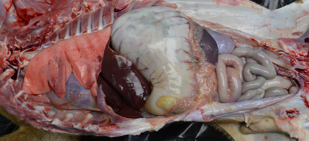
General view of the abdominal and thoracic cavities
The spleen and liver are separated from the digestive system to evaluate their shape, size, color, consistency and cut surface. In addition, the gallbladder (if it is full or empty, its perfusion, content, etc.) and the periportal lymph nodes will be assessed.
The digestive tract, is reserved for its opening and inspection at the end of the necropsy, although the pancreas and mesenteric lymph nodes will be visually evaluated at this point.
The adrenal glands will be examined in situ by means of a transversal section that allows us to evaluate the cortex-medulla-cortex ratio (1:2:1).
The genitourinary systems are then examined after its extraction from the abdominal cavity by cutting the dorso-lateral insertions to the abdominal cavity.
The kidneys are cut longitudinally, if possible with a single knife cut. Then, they are decapsulated by retracting the capsule with the help of forceps. The difficulty of this process, and renal parenchyma affectation has to evaluated.
The ureters should at least be visualized and palpated. The urinary bladder is opened and its contents are observed, as well as the appearance of its mucosa. In addition, the urethra and the adnexal glands of the male (prostate, bulbourethral glands and seminal vesicles) are assessed. In females, the ovaries are assessed by longitudinal section and their surface is explored. The uterus and uterine horns are opened longitudinally to assess the presence of abnormal contents and the appearance of the mucosa. In addition, the vagina and vulva will be assessed.
Thoracic organs
Two incisions are made parallel to both branches of the mandible to extract the tongue ventrally and the oral cavity and tonsils are examined. The hyoid branches are cut and the tongue, larynx, trachea are removed by pulling backwards, lifting the lungs with the heart and the thoracic part of the aorta, removing the pleural adhesions at the level of the thoracic spine. It is very important not to damage the lymph nodes and mediastinal nodes, as these structures are of great importance. Together with the esophagus, the aorta and the cava are also cut. All the thoracic organs can now be removed for examination on a table.
The thyroid and parathyroid glands are located, examined and removed.
The abdominal lymphatic organs: thymus in the case of young animals, mediastinal and bronchial lymph nodes, as well as the mediastinum are assessed.
The tongue is examined by transverse sections. The esophagus is opened longitudinally to assess the contents and mucosa and, in addition, it is separated from the trachea to assess the serosa.
The respiratory tract is separated from the heart and great vessels.
For the exploration of the respiratory system, first the larynx and trachea should be assessed by means of a longitudinal cut using scissors, and then assessing the lungs by visual inspection, palpation and cutting of the parenchyma, following the path of the large bronchi.
The heart and great vessels are then assessed. The heart should be assessed externally before being separated from the respiratory tract to better carry out the inspection of the great vessels and associated structures. First, the pericardium is examined and the state of the epicardium is observed (color, presence of adhesions or hemorrhages and epicardial fat) to then proceed to open it and see its cavities. Once examined, the relative size of the atria and ventricles has to be determined. The procedure to open the right and left heart varies. On the one hand, the right heart is opened by cutting parallel to the interventricular septum, starting from the ventricle at the level of the pulmonary artery trunk to the atrium and continuing parallel to the interventricular septum. On the other hand, the left heart is opened by a longitudinal cut from the atrium to the ventricle and then cutting through the aorta. The myocardium should be incised at the level of the septum.
Cranial cavity: Opening, and brain extraction and examination
The head is separated at the level of the atlantooccipital joint with the help of a scalpel and knife.
It is placed on the table to begin to remove the skin and muscles of the skull in order to have easier access to the bones. The cranial cavity is opened by two parallel cuts from the occipital foramen towards the base of the zygomatic process of the temporal bone.
Once the cuts are made, the detached part of the bones is lifted to expose the brain. In situ observation to assess the possible gross alterations is carried out.
To extract the encephalon, the dura mater is cut and, tilting the head from front to back, the encephalic mass is detached together with the pineal gland.
Next, the color and thickness of the meninges and parenchyma are assessed, as well as the conformation of the convolutions. Changes in shape, cystic structures, abscesses and granulomas or other localized volume increases had to be recorded. It is advisable not to cut the brain and to preserve it in 10% formalin for about ten days in order to evaluate it by histological sections.
Examination of joints and muscles
In the muscles, it is recommended to make several incisions at random locations.
Regarding joints, it is advisable to examine at least 5 joints assessing the content and the articular surface. It is possible to take advantage and evaluate those joints that have been opened during the necropsy. It is recommended to first make a longitudinal cut of the skin, change the scalpel blade and then make a transverse cut of the interarticular space to open the cavity and observe the appearance of the synovial fluid, the appearance of the articular surface and the synovial membrane.
Examination of the digestive tract
Finally, the examination of the digestive tract take place. The stomach and the pre-stomachs (in the case of ruminants), once their surface has been assessed, should be opened by the greater curvature and the intestine longitudinally along its entire length to be able to observe the mucosa, the food content and the serosa.
Sometimes it may be necessary to examine other parts such as:
- Nasal turbinates in the case of pigs to determine the possible existence of atrophic rhinitis and in the case of sheep for the presence of parasites such as Oestrus ovis.
- Spine in case of nervous symptomatology such as hemiplegia in the case of horses and dogs, as well as bone marrow in viral or neoplastic processes in small animals.
