Necropsy techniques
Regarding the methodology, there is a varied number of necropsy techniques described by different authors.
It is up to each individual pathologist to decide which of them to apply depending on his or her predilection for one or the other. However, for pathology students it is advisable to work following a specific necropsy protocol without great variation, in order to favor the fixation of concepts.
In any case, it is essential to always keep in mind that each case will require certain modifications depending on two fundamental factors:- The ante mortemdiagnosis: it will slightly orient the pathologist's search for lesions towards a certain specific organ or tissue.
- The diversity of species: the varied number of species to be studied in veterinary medicine means that the procedures are slightly modified according to the anatomical particularities of each species.
Although the technique of inspection of the internal organs is similar in all species, the positioning of the animals at the time of necropsy is very variable, although the right lateral recumbence position is recommended in all of them.
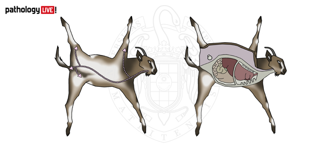
Thus the carcass of ruminants is placed in left lateral recumbence, to observe without problems the different abdominal organs avoiding the exit of the rumen.

Necropsy in the goat. Positioning in left lateral decubitus. Skin incision lines and opening of the abdominal and thoracic cavities.
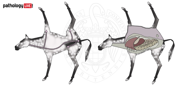
On the other hand, in dogs, cats, pigs and horses, a more orthodox positioning is preferred, in right lateral recumbence.

Necropsy in the horse. Positioning in right lateral decubitus. Skin incision lines and opening of the abdominal and thoracic cavities.
On the other hand, in the rest of species (mainly laboratory and wild animals) the preferred position is supine recumbence, which allows a complete inspection of all the abdominal organs at a glance.
